Radiology Continuing Education Series
Course 1 of 6
Physics of Radiology
Course 1 – Physics of Radiology
Course 2 – Choosing the Appropriate Exposure Factors
Course 3 – Recording the Image
Course 4 – Poor Quality Films-Causes and Corrections
Course 5 – Radiation Safety-Importance and Procedures
Course 6 – Pros and Cons of Digital Radiography-CR vs. DR
High quality x-rays first begins with understanding the nature and behavior of x-rays. X-rays are a form of electromagnetic radiation that travels in waves. Compared to visible light x-rays have a much shorter wavelength which allows them to penetrate materials that absorb or reflect visible light. The shorter the wavelength, the higher the frequency and penetrating power of the x-ray. All radiant energy travels in a straight line. The direction can be changed but the new path will also  continue in a straight line. The x-ray tube controls the direction of the x-ray beam.
How is the x-ray beam created?
X-rays are produced when fast moving electrons collide with a metal target. The energy carried in the electrons is converted to x-rays and heat. Only 1% of the energy is converted to x-ray energy and 99% into heat with temperatures in excess of 1832 degrees Fahrenheit.
How do you control the production of the x-rays and what characteristics of the x-ray beam can be controlled?
The operator can control: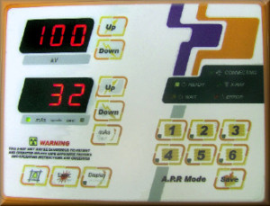
• The number of x-rays produced.
• The energy of the x-ray beam.
These controls are achieved through the exposure selections made on the x-ray machine operator console.
Knobology
kVp Selector: kVp stands for kilovolt peak/kilovolt potential. It affects the following:
• Selects the voltage differential used to accelerate the electrons.
• Gives variable “speed” to the electrons.
• Selects the penetrating quality of the x-ray beam.
• Effects the efficiency of x-ray production.
• Determines the scale of contrast in the image.
• Determines the scatter radiation produced by the patient.
Because x-ray production is basically the conversion of one type of energy, Kinetic energy, into two other types of energy, x-rays and heat, the energy of the x-ray is dependent on the energy of the electron at the time of conversion. The more energetic the electron is, the more energetic the x-ray will be. The faster the electron is moving, the greater it’s kinetic energy. The electrons kinetic energy is increased by implying an electrical potential between the cathode and anode of the x-ray tube. The  stronger this potential difference the greater the developed kinetic energy of the electrons.
 When you change the kVp you are changing the ability of the x-rays to pass through the object. Thicker and/or denser objects require more energetic x-rays to pass through them. Thinner and/or less dense objects require less energetic x-rays to pass through them.
The x-ray image is formed by the differential absorption of the x-ray beam. What you want to depict in your radiographs are the differences in the body’s tissues. If there are a lot of small differences you want to demonstrate that just as well as when there are big differences. The most common way of referring to differences is contrast. The range of 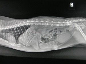 differences is typically referred to as a scale of contrast. When there are only one or two big differences this is referred to as a short scale of contrast. Controversially where there are a lot of small differences it is referred to as a Log scale of contrast. Obviously you need a kVp that is sufficient to penetrate the body area of interest. The lower the kVp, the more likely the x-ray is to be absorbed by most any type of tissue. At quite high kVp, the energy is sufficient enough that just about any tissue will be penetrated. So the objective of kVp selection is to choose a selection so that there is sufficient penetration, but not so great as to over-penetrate. Over penetration will not allow the differences or contrast to be depicted.
mA Selector: mA stands for milliamp ere: This is the measure of the number of electrons flowing in the x-ray tube.
• Activates a current flowing through the filament.
• Heats the filament causing electrons to be “boiled off”.
• Determines the number of x-rays produced per unit time.
• The number of x-rays reaching the film determines the degree of blackening of the film.
Exposure Timer: Controls the duration of electron flow being subjected to the voltage differential.
mAs Selector: mAs stands for milliampere seconds. It is the multiplication of mA by seconds, equaling mAs.
• This combines the functions of the mA selector and the exposure timer.
• Determines the total number of x-rays produced.
When you change the mAs you change the number of x-rays produced. Film blackening is directly proportional to the number of x-rays that pass through the object, room air or patient tissue.
Summary:
• kVp controls x-ray energy and thus penetrating ability and scale of contrast.
• mA x Seconds controls the total number of x-rays produced and thus film blackening.
• The most common failure of x-ray tubes is the result of technical error. To prevent costly damage to the x-ray tube it is important to use the appropriate kVp and mAs exposure selections to prevent damage to the anode.